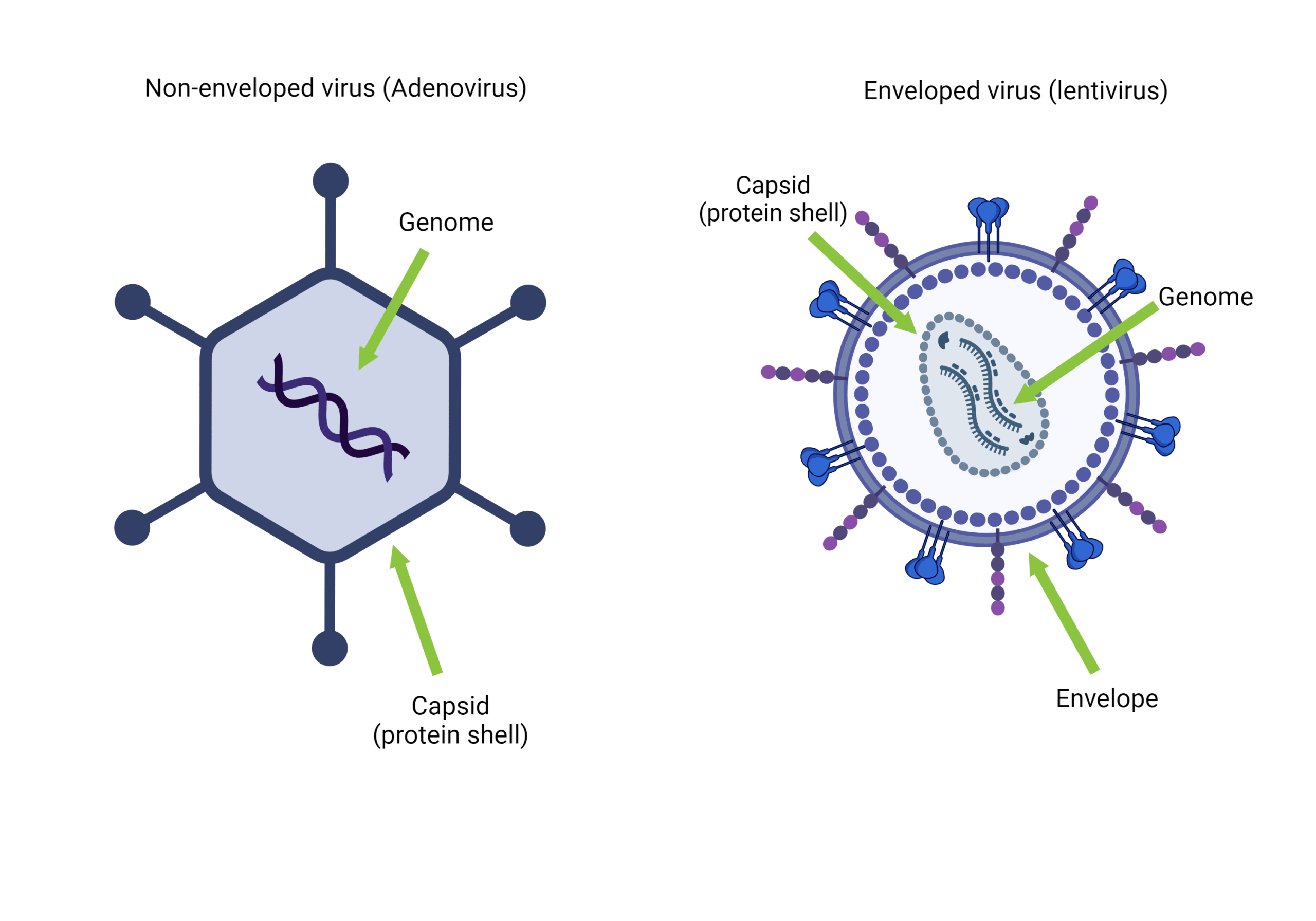
Due to its development as a hybrid of filamentous phage and plasmids, a phagemid is a cloning vector that can grow as a plasmid and be packaged as single-stranded DNA in viral proteins. Phagemid vectors differ from commonly used plasmids in that they can be loaded into the capsid of phages because they contain a phage packaging region of DNA from the genome of the phage. When compared to other vectors, phagemids have several advantages, such as producing single-stranded DNA that is ideal for sequencing and being easily used in site-directed mutagenesis.
Structure of Phagemids
A phagemid can be replicated as a normal plasmid expression vector and packaged in viral particles as single-stranded DNA. Phagemids have a double-stranded plasmid replication origin (ori) as well as an f1 ori for single-stranded replication and packaging into virus particles. Many popularly used plasmids have an f1 ori and thus are phagemids. They also contain the intergenic region of filamentous phages, which allows phagemid replication via helper phages and phagemid single-strand DNA packing. The selective marker region allows the selection of transformants, and the promoter site is responsible for directing the start of multiple cloning sites (where signal peptide, restriction sites, and tag are) while the coat protein site code for the virion capsid packaging.
Phagemid introduction into the cell
A phagemid can be introduced into a cell like a plasmid vector by electroporation and transformation just to mention a few. Also, combination with a ‘helper’ phage, such as VCSM13 or M13K07, is a unique way that phagemids can be introduced to a cell via a phage infection. The helper virion supplies the viral components required for single-stranded DNA replication and phagemid DNA packaging into phage particles. The helper particle, like a typical filamentous phage, infects the bacterial host by first attaching to the pilus of the host cell and then transporting the phage genome into the host cell’s cytoplasm. The virion genome causes the cytoplasm to produce single-stranded phagemid DNA within the host cell. The phagemid DNA is then packaged into phage particles. The bacterial host cell releases phage particles containing single-stranded DNA into the extracellular environment.
Filamentous phages partially inhibit bacterial growth but, unlike the lambda and T7 phages, are not typically lytic. Helper phages are ordinarily designed to package in a less efficient way (via a flawed phage ori) than the phagemid, resulting in phage particles that are composed primarily of phagemid DNA. Filamentous phage infection necessitates the presence of a pilus, so only microbial specifically bacterial hosts comprising the F-plasmid or its variants can generate phage particles.
Uses of phagemids
- Are useful for making templates for site-directed mutagenesis today (in-vitro mutagenesis).
- Can be used to generate single-stranded DNA templates for sequencing (which happened before the development of cycle sequencing).
- The detailed study of the filamentous phage life cycle and structural features resulted in the development of phage display technology, which allows a variety of peptides and proteins to be expressed as hybrids to phage coat proteins and displayed on the viral surface.
- Because the displayed peptides and polypeptides are linked to the respective coding DNA within the phage particle, this technique is useful for studying protein-protein interactions as well as other ligand/receptor combinations.


Leave a Reply
You must be logged in to post a comment.