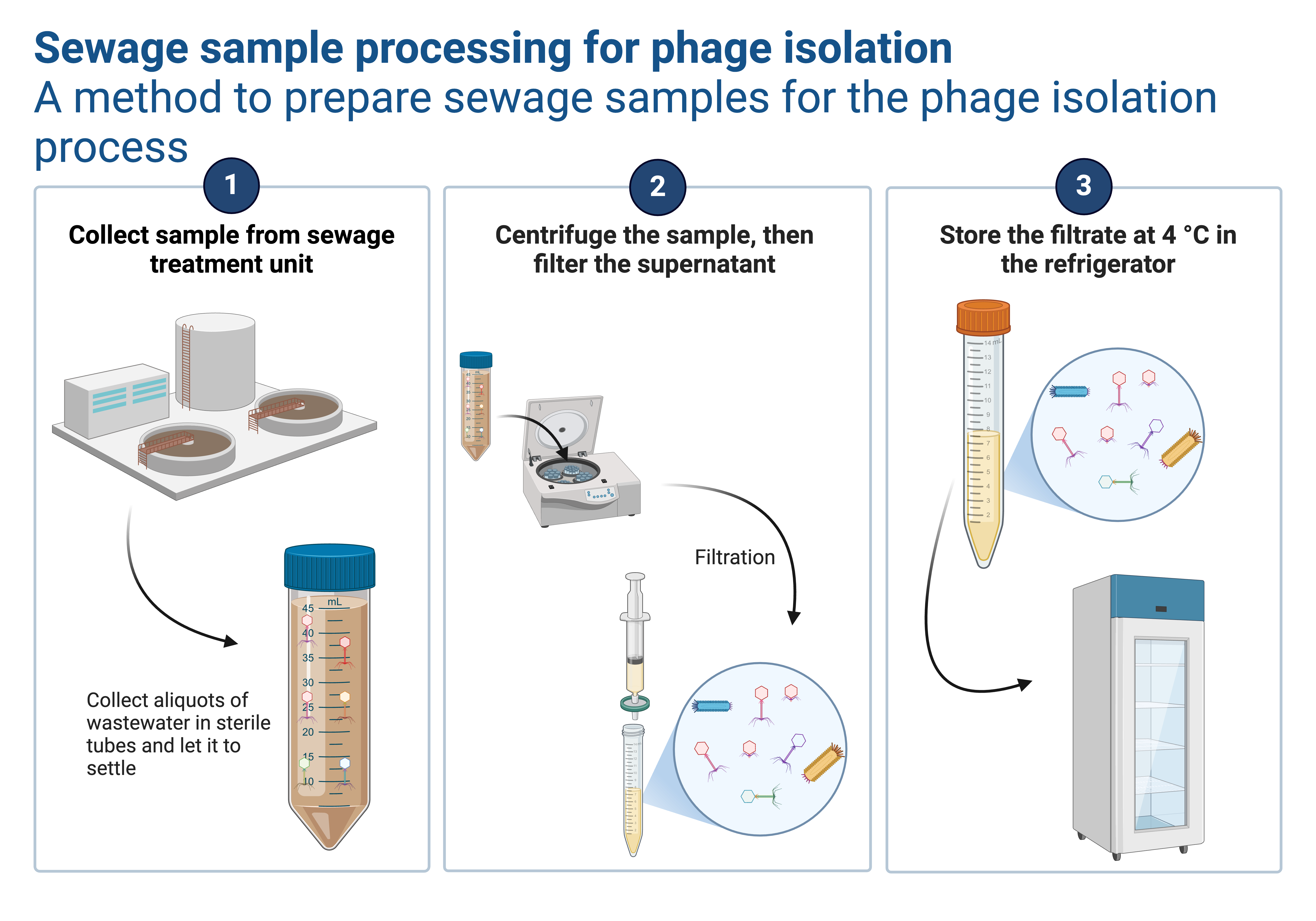
Bacteriophages are viruses that are particularly reliant on their bacterial hosts’ metabolic activity to copy their genetic material and produce the proteins necessary for their survival. They are showing potential antibacterial effects against a small selection of dangerous microorganisms. A bacteriophage infects a host cell by attaching to a bacterial strain that is vulnerable to it. The infection either causes the cell to lyse or causes the viral genome to merge with the host genome, processes known as lytic or lysogenic replication cycles. Phages with lytic replication cycles are used in a variety of situations where the goal is to kill microorganisms.
Phages only infect one species or even one host strain and have a narrow range of hosts. This suggests that unique therapeutic formulations are required to focus on the virus that is causing the sickness. Therapeutic phage preparations frequently contain a variety of distinct phages to broaden the host range and overcome any possible resistance. Selecting the ideal phage combination for anti-bacterial therapy can be challenging. Thus, methods that let us compare the activity of different phages are essential.
Phages are able to adhere to various replication cycles. The term “temperate phage” refers to phages that have the ability to lysogenize or enfold their genome within the chromosome of their bacterial host. Phages employed in therapy, on the other hand, are often obligately lytic. After invading the host, these phages multiply and create new virus particles by utilizing the host’s cellular machinery. The new phages will burst out of the host cell, causing it to lyse, after enough progeny have been created.
Ways of quantifying Bacteriophages and their limitations
Finding phages that can infect the target pathogen is a crucial stage in the phage therapy process. Traditionally, phage suspensions are spotted on a bacterial lawn in a plaque assay to accomplish this. A distinct region known as plaque emerges in the bacterial lawn where the phages are inhibiting or lysing bacterial development. Double-layer agar (DLA) experiment is a version of this technique that involves combining bacteria and phages in soft agar and then covering it with a solid agar plate. By counting the individual plaques that are produced in both situations, the number of infective phages as plaque-producing units can be ascertained. These traditional methods are popular and inexpensive, they can only provide results after overnight incubation and cannot evaluate the dynamics of phage infection. Additionally, they are difficult to scale up for high-throughput screening and are not always precise or reproducible.
To circumvent these limitations, various approaches for measuring bacteriophage killing have been developed. One of these is to measure the optical density of a bacterial culture and watch how it changes over time as the phage infection grows. Although this form of assay does provide in-situ infection monitoring, it merely evaluates the turbidity of the bacterial culture, constituting an oblique measurement of bacterial damage. Techniques based on bacterial respiration have also been researched. A one-step growth curve is an additional test that could be utilized. In this method, the host bacteria and the phages are incubated together, and aliquots of the free phage are taken at various time points and plated on double-layer agar. This test is regarded as the gold standard for phage characterization. It can be used to calculate their latent period, or the number of new phages created per bacterium, as well as their generation rate (burst size). But like the DLA assay, this assay requires a lot of labor and is challenging to scale up. Phage infections can be tracked in a multi-well plate environment in real-time. As the phage infection develops, sytox green, a membrane-impermeant nucleic acid dye, stains the DNA of lysed bacteria and nascent phage progeny and emits a fluorescent signal.
References
Egido, J. E., Toner-Bartelds, C., Costa, A. R., Brouns, S. J. J., Rooijakkers, S. H. M., Bardoel, B. W., & Haas, P. J. (2023). Monitoring phage-induced lysis of gram-negatives in real-time using a fluorescent DNA dye. Scientific Reports, 13(1), 1–12. https://doi.org/10.1038/s41598-023-27734-w


Leave a Reply
You must be logged in to post a comment.