Bacteriophage sample processing

Sewage sample processing for bacteriophage isolation

In our previously published article focusing on sewage sample collection, we explored the presence of bacteriophages in the environment. Sewage stands out as a readily accessible source with abundant concentrations and diverse types of bacteriophages. Notably, Coliphages, which are viruses that target coliform bacteria, can be found in sewage wherever coliforms are present, typically ranging from 10^5 to 10^7 per liter.

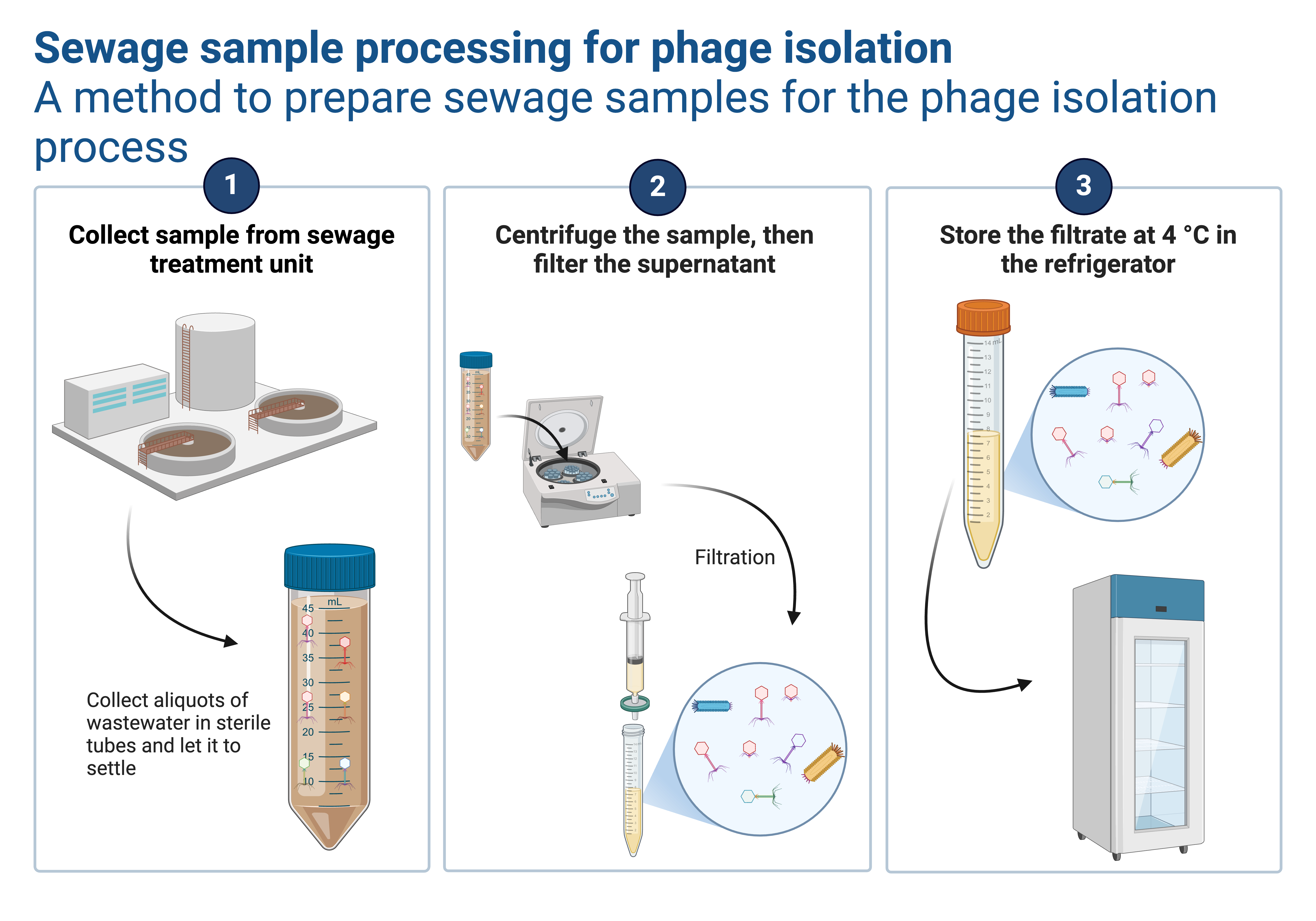
Similar to handling any other laboratory sample, the collected sewage sample requires specific processing procedures for bacteriophage isolation. This article aims to provide a detailed, step-by-step guide on how to process a sewage sample to successfully isolate bacteriophages.

Processing steps

Preprocessing

Once the sample is collected, it must undergo several preprocessing steps to remove debris and concentrate the bacteriophages:

  1. Allow the sample to settle for a brief period to allow larger particles to settle to the bottom.
  2. Carefully decant the supernatant, leaving behind the settled particles.
  3. Filter the supernatant using a sterile filter with a pore size small enough to retain bacteria but large enough to allow phages to pass through.
  4. Concentrate the filtered liquid using centrifugation or ultrafiltration techniques.

Bacterial Host Preparation

To successfully isolate bacteriophages, it is essential to cultivate specific bacterial strains that can act as hosts for the phages. Follow these steps to prepare the bacterial host:

  1. Choose a bacterial strain known to be susceptible to phage infection and relevant to your research objectives.
  2. Cultivate the bacterial host using appropriate growth media and incubation conditions.
  3. Verify the purity of the bacterial culture to avoid contamination.

Phage Isolation

With the sample preprocessed and the bacterial host prepared, you can proceed to isolate bacteriophages from the sewage sample:

  1. Mix the concentrated sewage sample with the bacterial host culture.
  2. Allow the mixture to incubate for several hours to allow phage infection and replication to occur.
  3. After incubation, perform plaque assays or other techniques to identify areas where phage infection has caused bacterial cell lysis, resulting in clear zones or plaques.
  4. Carefully pick individual plaques using a sterile pipette tip and transfer them to separate tubes containing suitable growth media to establish isolated phage cultures.

Bacteriophage isolation in details

Requirements
  • Host bacteria such as Escherichia coli broth culture (3-5 hrs)
  • Soft agar (top agar- nutrient agar with 0.7% agar)
  • Nutrient agar plates (also TSA and LBA can be used)
  • Sterile 1ml pipettes
  • 9.0ml buffered saline blanks
  • Water bath (50°C) and incubator at 37°C.
The procedure of isolation of bacteriophages
  1. Dilute the sewage sample at 1:10 and 1:100 in the buffered saline blank.
  2. Cool under running water four tubes containing sterile soft agar (3ml/tube) to 50°C. Label them as 1,2,3,4.
  3. Aseptically add 1ml of undiluted sewage in each tube and Host bacteria young culture to tube 1 and mix the tube contents thoroughly. Pour it immediately onto the surface of dried nutrient agar and let it spread uniformly over the entire surface by rotating the Petri plate.
  4. Repeat the experiment by adding 1ml diluted sewage (1:10 and 1:100) to tubes 2 & 3 and mixing it with 1ml bacterial culture. Treat control tube 4 similarly but add buffered saline in lieu of sewage sample.
  5. Let the agar solidify. Invert the plates and incubate at 37°C (incubation may change depending on the host used) for 4hrs.
  6. At the end of incubation, count the number of plaques in each dilution and calculate the concentration of phages in the sewage sample. Record the size and shape of plaques.

Phage Characterization

Once isolated, the bacteriophages should undergo a comprehensive characterization process to understand their properties and potential applications:

  1. Perform morphological analysis using electron microscopy to determine the phage’s structure, shape, and size.
  2. Conduct genomic analysis through DNA or RNA extraction, followed by sequencing and bioinformatics analysis to identify the phage’s genetic makeup.
  3. Assess the phage’s infectivity range by testing it against different bacterial strains.
  4. Evaluate the phage’s lytic or lysogenic activity to understand its behavior and potential applications.

The isolation and characterization of bacteriophages from sewage samples offer significant scientific and practical value. By following the step-by-step guide outlined in this article, researchers can successfully process sewage samples and isolate bacteriophages for further study. These efforts can lead to new insights into phage biology, potential therapeutic applications, and their role in environmental processes. With continued research in this field, bacteriophages may prove to be powerful tools in combating antibiotic resistance and addressing various challenges in medicine and beyond.

About the author

Hello there!

I'm Raphael Hans Lwesya. I have a deep interest in phage research and science communication. I strive to simplify complex ideas and present the latest phage-related research in an easy-to-digest format. Thank you for visiting The Phage blog. If you have any questions or suggestions, please feel free to leave a comment or contact me at [email protected].

Comments

  1. I do not even know how I ended up here, but I thought this post was great.
    I don’t know who you are but certainly you are
    going to a famous blogger if you are not already 😉 Cheers!

Leave a Reply

Receive the latest news

Subscribe To Our Weekly Newsletter

Get notified about new articles